Visco-elastic Behavior of Cells in a Microfluidic Device using FSI
Variations of structure and shape of cells play an important physiological role. For instance, tumor and normal cells can be distinguished by elasticity, indicated by the amount of deformation under given stress. The mechanical characterization of a certain cell type is meaningful to obtain (patho-) physiological insight. Simulations help to understand, verify and improve the analysis of deformation-based cell characterization such as CAOS [1] or flow-based cytometry [2].
We aim to provide a simulation-based database for the mechanical deformation of cells in microfluidic channels. The variation of parameters of the viscoelastic models for the cells results in a library of possible cell deformation classes.
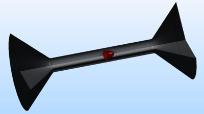
A cell develops characteristic shapes, while moving through a microfluidic channel with varying width. We achieve efficient computations using a 2D-rotational symmetric model, based on Fluid-Structure-Interaction with a hyper-elastic material. The influence of different parameters on the deformation is evaluated and will be presented. The deformation of a cell along an entire microfluidic channel can be tracked for a variety of elasticities, viscosities and flow rates. This research allowed to create guidelines for channel geometries specific for certain cell types or particles.
[1] Tobias Neckernuss et al., Setup and analysis to stretch adherent cells with light, Proc. SPIE 10876, Optical Interactions with Tissue and Cells XXX, 1087607 (1 March 2019); doi: 10.1117/12.2508475;
[2] Otto et al., Real-time deformability cytometry: on-the-fly cell mechanical phenotyping, Nat. Methods, 2015
Download
- CC_Cambridge19_Ralf.pptx - 1.45MB